Health Going Digital
Not so long ago, say 60 years, dentists or so-called barber dentists used to extract teeth and place them in a showcase outside their clinic to lure potential patients requiring difficult extractions. A candle would illuminate the extracted bloodstained tooth and a sign would welcome them in. Gone are the days of barbaric dentistry, in comes the age of digital dentistry.
Every profession owes most of its advances to computers. This is the same with dentistry. Conventional methods are fast dieing out, being replaced by new digital techniques.
Areas of digital Dentistry:
- CAD/CAM and intraoral imaging — both laboratory- and clinician-controlled
- Caries diagnosis
- Computer-aided implant dentistry — including design and fabrication of surgical guides
- Digital radiography — intra oral and extra oral, including cone beam computed tomography (CBCT)
- Lasers
- Shade matching
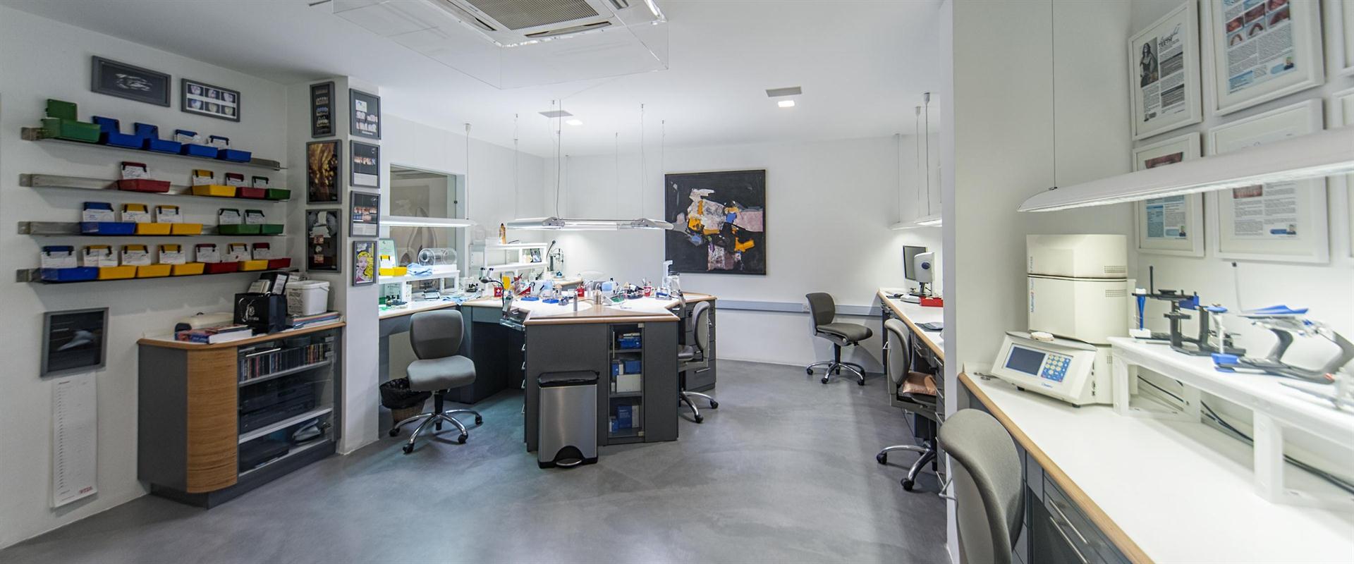
A large field of digital dentistry lies within CAD CAM or computer-aided dentistry and computer-aided manufacturing. This encapsulates the production of crowns and bridges as well as allows for the fabrication of surgical guides aiding implant placement. Clinical dental chair-side CAD CAM equipment (figure 1) is used to take digital photos of your teeth. Thousands of these photos shot in seconds are saved together giving a 3-Dimentional image of the patient’s teeth. With the help of specific dental software, these images are then used to design the crowns and bridges. At this stage patients sitting in the dental chair may view the design and request some modifications. The length, width and angulation of the designed teeth are some of the modifications patient may wish for so in order to achieve the set of teeth they always desired. Once the design is agreed on, the type of material is chosen.
Figure 1: A chair-side CAD CAM unit (pic to be sent in another email)
The material comes in pre-fabricated blocks of conventional metal-ceramic or all-ceramic metal-free blocks. The data is then sent by wifi to a milling unit (Figure 2). The milling unit cuts or mills the block into the designed tooth saved on the software. In this manner the crown or bridge is ready in minutes. This method of delivery allows for the same day/appointment service of completing ones crowns (Figure 3).
Figure 2: A CAD CAM milling unit (pic to be sent in another email)
Figure 3: A milled all-ceramic metal-free crown (pic to be sent in another email)
With the introduction of cone beam computational tomography or CBCT scans, radiology has also benefited greatly. As opposed to conventional CT scans, this 3D data is acquired using lower radiation doses more specific to dentistry. Using the software (Figure 4), the data is used to design and produce a surgical guide (Figure 5). The surgical guide is used to place implants without the need of incising and suturing the gums up as in conventional implant dentistry. The advantages are great! Higher precision, close to no bruising and improved overall results are but a few to mention.
Figure 4: Software used to produce a surgical guide (pic to be sent in another email)
Figure 5: The surgical guide (pic to be sent in another email)
Digital dentistry is highly advantageous to the patient as it offers them quick, custom-made patient-specific solutions using only the latest in dental technology. Ask your dentist what’s out there for you!